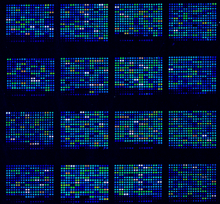
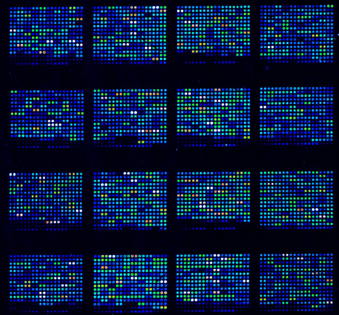
Microarray technology is one of the most sophisticated methods used by the scientific world. Scientists make use of the principal of DNA microarray technology and utilize it in hybridizing immobilized DNA molecules. This is a complex, three part step that can help scientists and researchers to achieve the process.
-
Manufacturing – This step requires the availability of a chip or microarray. It is a simple glass or silicon slide that is manufactured by complex robotics and is given a special surface that is used to make microarrays.
-
Sample preparation and array hybridization – To achieve this, the DNA and miRNA have to be isolated, and then followed up by the labelling of cDNA probes to enable the mobilization of the targeted DNA.
-
Data analysis – The final step is scanning and image analysis conducted by the scientist and researcher. Using highly sophisticated software, various programs allow researchers to interpret the collected data.
Basically, a microarray experiment involves the comparison of a question or a sample that represents all the genes being analysed. A simple example of this is the study of bacteria being bred in an infected cell, while simultaneously being grown in standardized, controlled atmospheres. Similar comparisons will show the difference in the specimens, thereby allowing researchers to find a cure and to develop medications when they collaborate with doctors.

A basic microarray experiment involves four steps. The steps are as follows:
1. Preparing a Sample – The sample preparation starts by isolating a RNA messenger that represents the quantitative copy of genes expressed when the sample was collected. This step is crucial because the entire process depends upon the quality of the RNA taken for the microarray experiment.
2. Hybridization – Hybridization is a process through which two complementary strands of DNA are joined to form a double stranded molecule. Here, the Sample and Control DNA are mixed together, then purified by removing elements like lipids, carbohydrates, proteins, unincorporated nucleotides and primers. Before hybridization, the microarray slides are incubated at high temperatures with solutions like SSC (saline-sodium buffer), SDS (Sodium Dodecyl Sulfate) and BSA (bovine serum albumin), so that it can reduce its background.
3. Cleansing – The slides are then washed after the hybridization so that the cDNA can be removed and thus remained untouched by the hybridization. Moreover, cleansing increases the severity of the experiment to reduce cross hybridization.
4. Analysis – Image acquisition and data analysis is the last stage of the experiment. The aim is to produce an image of the hybridized array. The slide is dried and placed on a laser scanner to observe the results.
microarrays can be applied in a wide array of sectors. They help everything from disease monitoring to eradicating signs of illnesses from the cells. They allow doctors and researchers to work together, so that they can provide a better service to the beneficiaries of the medical world. With advancements in technology, there is little left unseen by scientists. Their printers and tools all help in gaining a better understanding of how molecules and genes work.
Rebecca Craig is one of the leading experts in genomic research. She has worked with microarrays, and often read and write on the subject. She suggests this website, to anyone looking for microarray printing services.


